Committees / Technical Committee/Newsletter/11.06.20 april2
01.01.1970
 01
01
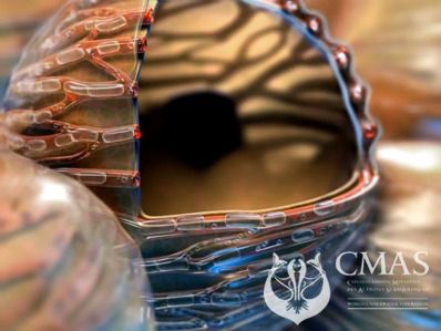
Given that micro-bubbles (MBs) obstructs blood vessels and ascents cause MBs (or so-called free-gas), it would seem common sense to slow the rate of ascent and include DeeP-Stops!
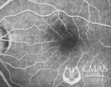
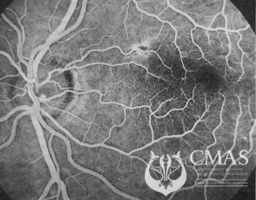
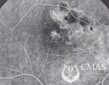
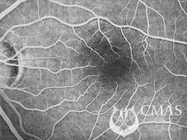
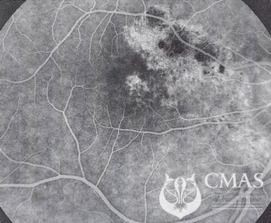
Keep in mind that the eyes are “end organs” of the brain and are a window into the brain that needs no scalpel. Capillary damage seen here is more than likely to be mirrored in the brain itself.
 01 01 |
 01 01 |
 01 01 |
| A Normal Retina | Minor Retina Damage | Gross Retina Damage |
Ear Clearing?
Think on! You may have a PFO without knowing it - about 25% of us do!!. Remember that clearing your ear using Valsalva’s maneuver may shunt MBs into your arterial circulation - DCI may be next!. Find another way to clear your ears and add DeeP-Stops into your dive profile - please.
Safe Diving
Bob Cole
CMAS Technical Director


